
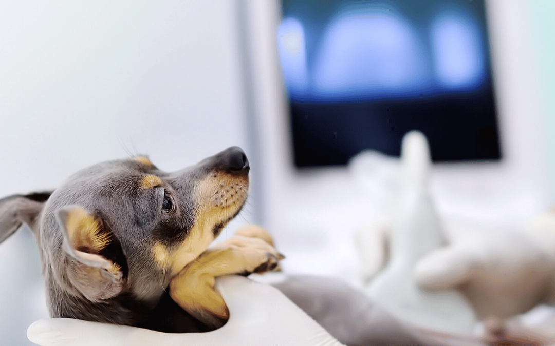
Advanced imaging allows your pet’s veterinary team to look beyond what can be seen or felt during a physical exam. These tools provide detailed views of soft tissues, organs, bones, foreign objects, and internal structures—revealing conditions that may otherwise go undetected.
Below is an overview of the advanced diagnostic and imaging modalities we offer, how they work, and when your pet might benefit from each one.
Radiography (X-Rays)
Radiography is one of the most commonly used diagnostic imaging tools in veterinary medicine. Using controlled X-ray beams, radiographs create two-dimensional images that are especially effective for evaluating bones, the chest cavity, and abdominal structures.
Radiography is commonly used to assess:
- Bone fractures and joint abnormalities
- Arthritis and degenerative joint disease
- Chest conditions affecting the lungs or heart
- Abdominal organ size and positioning
- Gastrointestinal obstructions or foreign bodies
- Bladder stones
Radiographs are fast, widely accessible, and often serve as the first diagnostic step in evaluating illness or injury. While highly valuable, radiography provides less detail for soft tissues compared to ultrasound or CT.
In simple terms: X-rays provide a quick snapshot of internal structures, especially bones and the chest.
CT (Computed Tomography)
CT imaging combines X-ray technology with advanced computer reconstruction to create detailed, cross-sectional images of the body. This method excels at visualizing bones and complex anatomy and produces three-dimensional images quickly and efficiently.
CT imaging is commonly used for:
- Complex orthopedic injuries or fractures
- Head and neck trauma
- Nasal disease
- Dental disease and impacted teeth
- Tumor staging
- Joint evaluation
- Pre-surgical and radiation therapy planning
- Detailed organ assessment
- Vascular evaluation (with contrast)
CT scans offer excellent anatomical detail and rapid image acquisition, making them especially valuable in urgent or complex cases. While highly advanced, CT provides less soft-tissue contrast than MRI.
In simple terms: CT creates detailed “slice-by-slice” images of the body, allowing veterinarians to view bones and soft tissues in three dimensions.
Ultrasound
Ultrasound uses high-frequency sound waves to generate real-time images of internal structures, making it an excellent tool for evaluating soft tissues and organ function.
Ultrasound is commonly used to assess:
Abdominal Structures
- Liver, spleen, and kidneys
- Gastrointestinal tract and wall layering
- Bladder and ureters
Cardiac Evaluation (Echocardiography)
- Heart chamber size and function
- Heart valve structure and performance
Guided Procedures
- Biopsies
- Fluid aspiration
- Catheter placement
Ultrasound provides dynamic, real-time imaging without radiation exposure and allows for precise, guided sampling. Limitations include reduced image quality when bones or gas obstruct the view and decreased effectiveness for deep or thoracic structures without appropriate acoustic windows.
Advanced Diagnostics, Thoughtfully Applied
Each imaging modality serves a distinct purpose. Your veterinarian will recommend the most appropriate diagnostic tool based on your pet’s symptoms, medical history, and clinical needs—ensuring accurate diagnosis while prioritizing comfort and safety.
If you have questions about any imaging recommendation or would like help understanding which diagnostic option is right for your pet, our team is always here to guide you through the process.