
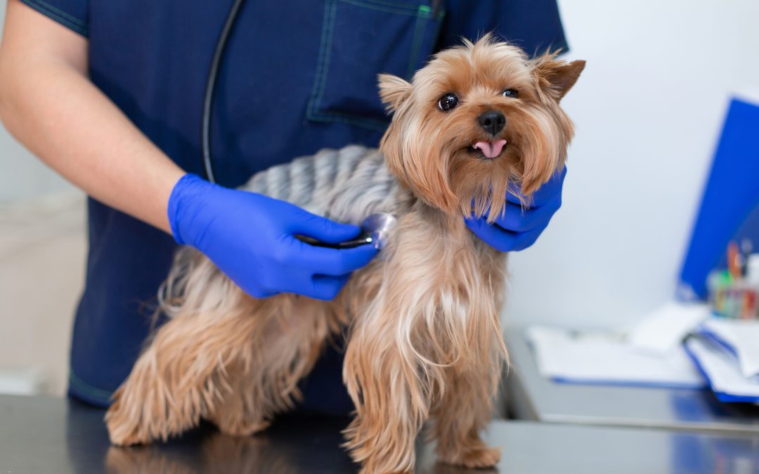
Veterinary cardiology focuses on the structure and function of the heart and blood vessels, with the goal of diagnosing and treating cardiovascular disease in pets. Much like human cardiology, veterinarians use advanced diagnostic tools such as echocardiograms and electrocardiograms (ECGs) to identify and manage conditions including heart failure, arrhythmias, and valve disorders.
Cardiac procedures range from low-risk diagnostic tests to higher-risk interventional procedures, most of which are minimally invasive. Veterinary cardiologists may manage conditions such as leaky heart valves, abnormal blood flow (shunts), or fluid accumulation in the chest cavity, helping improve both quality of life and long-term outcomes for affected pets.
Echocardiography
Echocardiography is a noninvasive ultrasound examination of the heart and is considered the gold standard for diagnosing many forms of heart disease. This imaging allows real-time assessment of the heart’s size, structure, function, and blood flow. It is commonly used to diagnose conditions such as heart failure, congenital defects, heart tumors, and heartworm disease.
The procedure is performed using a handheld probe placed on the pet’s chest. The probe emits high-frequency sound waves that bounce off cardiac structures and return as echoes, which are converted into detailed images. Echocardiography is safe, painless, and highly informative, providing critical information needed for accurate diagnosis and treatment planning.
Electrocardiography (ECG)
Electrocardiography is a noninvasive method of recording the electrical activity of the heart using sensors placed on the skin. Also known as an EKG or ECG, this test evaluates heart rate, rhythm, and electrical conduction. It is essential for diagnosing arrhythmias, assessing chamber enlargement, and guiding anesthetic monitoring.
One limitation of ECGs is that they measure electrical activity rather than mechanical function, meaning they cannot directly assess how strongly the heart is pumping. For this reason, ECGs are often used alongside echocardiography for a complete cardiac evaluation.
Putting It All Together
Heart disease in pets can sound intimidating, but advances in veterinary cardiology allow for earlier diagnosis, more precise treatment planning, and improved long-term outcomes than ever before. With tools like echocardiography and electrocardiography, veterinarians can evaluate both the structure and electrical function of the heart to develop a comprehensive understanding of your pet’s condition.
If your pet has been diagnosed with a heart murmur, is experiencing coughing, exercise intolerance, fainting episodes, or other concerning symptoms, a cardiology evaluation can provide clarity and direction. Early assessment often leads to better management options and improved quality of life.
At TruVet Specialty & Emergency Hospital, our goal is to combine advanced cardiac diagnostics with clear communication and compassionate care — helping you feel informed and confident in every step of your pet’s treatment journey.
If you have questions about your pet’s heart health or would like to schedule a cardiology consultation, our team is here to help.